
From Ankle Sprain to the Spine
An ankle sprain looks like a local problem. This illustrative case shows, in seven steps, a hypothesis of how compensations can climb through the fascia up to the pelvis and the spine. Choose a phase to see it on repeat and read the explanation.
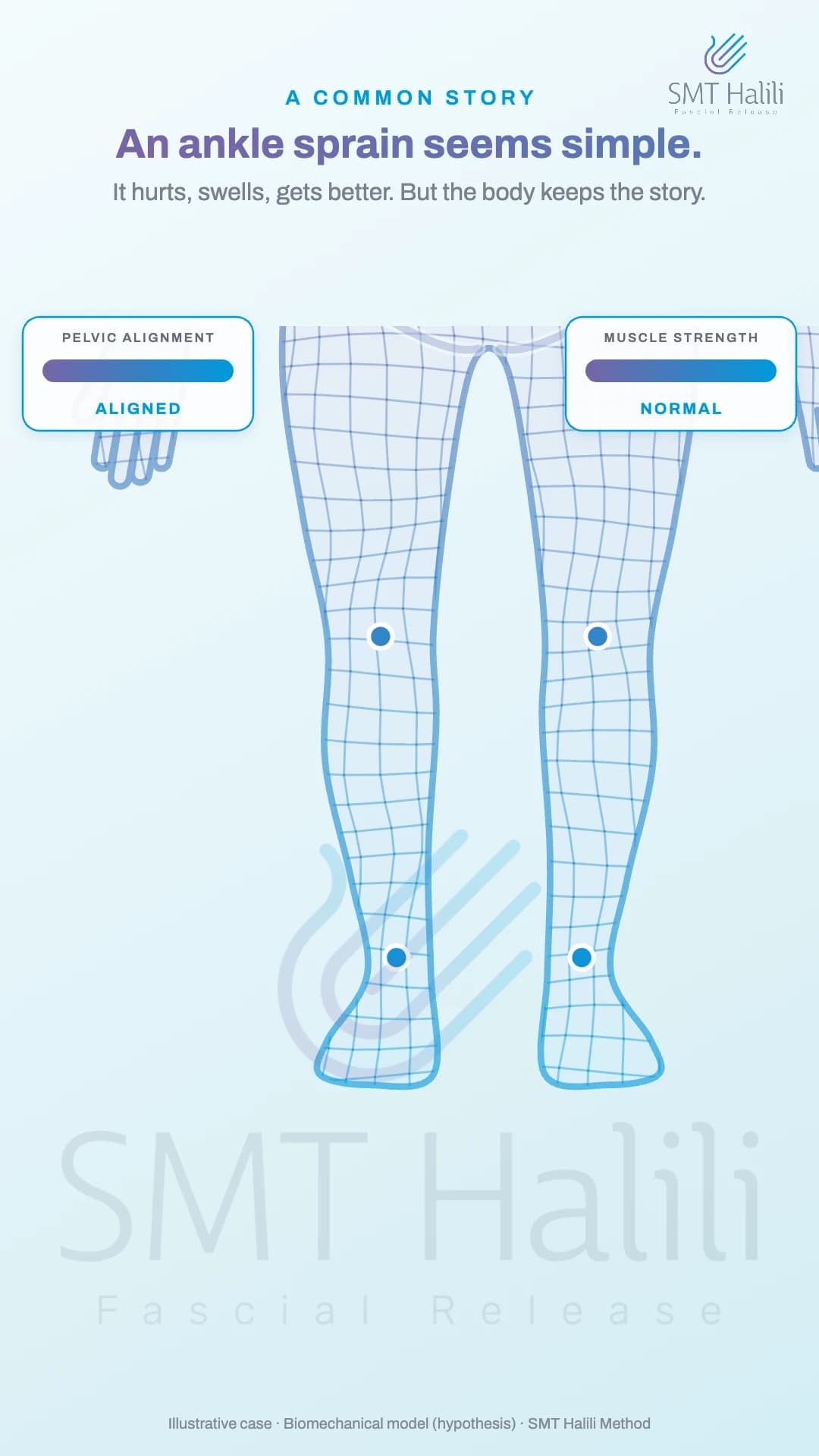
Phase 1 of 7
A COMMON STORY
An ankle sprain seems simple.
It hurts, swells, gets better. But the body keeps the story.
Learn more
An ankle sprain seems simple: it hurts, swells, and gets better. But the body keeps the story of that injury.
For professionals
Ankle sprain: a common injury, often considered resolved once pain and swelling subside. Illustrative case of a biomechanical model (hypothesis): each link has some isolated support in the literature, but the full sequence, from sprain to spine, has not been demonstrated in studies.
Phase 2 of 7
THE SPRAIN
Beyond the ligament, the nerve is stretched.
In inversion, the fibular nerve can be pulled. The central nervous system tells the fascia to guard the area.
Learn more
In a sprain, beyond the ligament, the nerve on the outside of the leg can be stretched. The central nervous system gets this alarm and tells the fascia to guard the area.
For professionals
Inversion sprain: beyond the ligament injury, the fibular nerve can be pulled. Reduced nerve conduction after the sprain and nerve strain in a cadaver model have been reported, more often in severe sprains (Nitz et al., 1985; Kleinrensink et al., 1994; O'Neill et al., 2007); low to moderate evidence. Central sensorimotor changes are described in chronic ankle instability (Hertel & Corbett, 2019; Maricot et al., 2023). “Fascia on guard” is the SMT method's reading of this protective response.
Phase 3 of 7
MUSCLES SWITCHED OFF
The stabilizers lose their command.
The fibularis muscles lose strength. The gluteus medius on the same side may weaken too.
Learn more
The muscles on the outside of the leg (fibularis) lose strength. The gluteus medius, a hip muscle on the same side, may weaken too.
For professionals
In chronic instability, reflex excitability of the fibularis muscles is reduced (arthrogenic muscle inhibition, small effect) and evertor strength is impaired (McVey et al., 2005; Kim et al., 2019; Khalaj et al., 2020); these findings do not appear in the tibialis anterior. Weaker hip abductors are common in chronic instability (DeJong et al., 2020; Zheng et al., 2025), including on the sprained side (Friel et al., 2006), but not in the first three months after a first sprain (Khalaj et al., 2022). The nervous system plays a part in the weakness; a reflex mechanism at the hip has not been demonstrated.
Phase 4 of 7
THE LATERAL CHAIN
Tension climbs through the fascia.
Without support, the foot supinates and the tibia rotates out. Tension follows the lateral chain up to the hip.
Learn more
Without that support, the foot turns inward and the leg rotates outward. In this model, tension climbs through the fascia on the outside of the leg up to the hip.
For professionals
Rearfoot and tibia move in a coupled way: calcaneal inversion goes with external tibial rotation, and injury to the anterior talofibular ligament alters tibiotalar rotation (Fischer et al., 2018; Caputo et al., 2009). Anatomical continuity between the leg fascia, the iliotibial tract, and the hip exists, but tension transmission along the lateral line has not been measured in living people (Wilke et al., 2016; Krause et al., 2016).
Phase 5 of 7
THE PELVIS
The pelvis loses its level.
It drops to the opposite side, the ilium rotates, the sacrum twists. The injured leg becomes “shorter”.
Learn more
In this model, the pelvis loses its level: it drops to the opposite side and rotates. The leg on the sprained side starts working as if it were shorter.
For professionals
Leg length differences alter pelvic obliquity and torsion (Betsch et al., 2012; Cooperstein & Lew, 2009), and the measurement of a “functional short leg” has uncertain reliability (Knutson, 2005). No study has linked ankle sprain to pelvic drop, iliac rotation, or sacral torsion: this link is a hypothesis of the model. In the clinical observation of the SMT method, a right ankle sprain usually comes with posterior rotation (retroversion) of the right hemipelvis, but the pattern varies with other restrictions in the patient's body.
Phase 6 of 7
THE S-CURVE
The gaze returns to level.
The spine compensates in an S. In this model, an old sprain may contribute to low back, neck or shoulder pain.
Learn more
To keep the eyes level, the spine compensates in an S shape. In this model, an old sprain may contribute to low back, neck, or shoulder pain.
For professionals
The spine compensates in the frontal plane when the length difference exceeds about 2 cm (Betsch et al., 2013); the double S-curve has not been measured. Ankle and lower-limb injuries are associated with more low back pain in large cohorts, without proof of causation (Seay et al., 2018; Foster et al., 2023). There are no studies on neck pain; for the shoulder, only indirect and very weak evidence (Leroux & Lagniaux, 2024).
Phase 7 of 7
FROM FOOT TO HEAD
That is why SMT looks at the whole chain.
The goal is for the fascia to glide again and the muscles to regain their command.
Learn more
That is why SMT looks at the whole chain, from foot to head. The goal is for the fascia to glide again and the muscles to regain their command.
For professionals
A whole-chain approach, not just the ankle. Treatment goal: restore fascial glide and muscle command. This is the clinical reasoning of the SMT method, not a demonstrated result for this illustrative case.