
SMT HaliliFascial Release

Fascia

Interwoven white fibers over a purple-to-blue gradient, suggesting the continuous network of fascia
Representation of fascia as a continuous network. AI-generated image, with no value as an anatomical reference.

Fascia is a connective tissue that is ubiquitous in the human body. It surrounds, interconnects, and supports every structure (muscles, bones, nerves, organs, and blood vessels), forming a continuous three-dimensional network. In the biotensegrity model (Levin & Martin, 2012), this network distributes tension and support throughout the body, which helps explain why a local restriction is rarely resolved by looking only at the spot that hurts.

Structure and properties

Made up predominantly of collagen and elastin fibers, fascia combines strength and elasticity. This allows it to bear loads, absorb impact, and facilitate mobility without compromising its structural integrity (Schleip et al., 2012; Stecco, 2015).
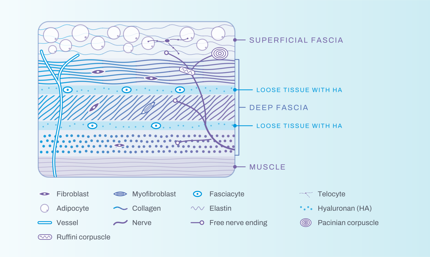
Layered diagram of fascia: superficial fascia with adipocytes; loose tissue with hyaluronan; deep fascia with collagen fibers in alternating directions; and muscle, crossed by vessels and nerves. The legend identifies fibroblast, myofibroblast, fasciacyte, telocyte, adipocyte, collagen, elastin, hyaluronan, vessel, nerve, free nerve ending, and Pacinian and Ruffini corpuscles.
Schematic illustration of the composition of fascia, from the superficial fascia to the muscle. No value as an anatomical reference.

It also has its own capacity for contraction, mediated by myofibroblasts, cells found even in healthy fascia that adjust the tissue's stiffness. Over minutes, this contraction can influence movement coordination; over days to months, it can lead to lasting tissue contractures (Schleip & Klingler, 2019).

A communication network

More than a structural link, fascia is a body-wide communication system. Its rich vascularization and innervation, together with the presence of mechanoreceptors and nerve endings, make it sensitive to a variety of stimuli. In this way, it relays information to the central nervous system, contributing to proprioception, balance, and the perception of the body in space (Findley & Shalwala, 2013; Stecco et al., 2016).
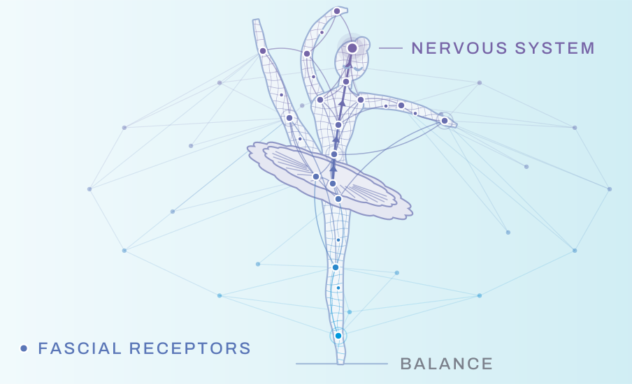
Schematic illustration of a ballerina balancing on one leg, the body wrapped in a mesh with points linked by lines that rise to the head; labels point to the nervous system, the fascial receptors, and balance
Schematic illustration of proprioception: fascial receptors inform the nervous system about position and balance. No value as an anatomical reference.

When fascia loses its health

Healthy fascia allows smooth movement: its layers glide over one another thanks to the hyaluronan between them. Tension, trauma, or changes in this lubrication can create restrictions, associated with myofascial pain and with impaired coordination, proprioception, and balance; in the superficial fascia, with symptoms of venous and lymphatic circulation (Stecco et al., 2011; Stecco et al., 2016). That is why fascial health is fundamental to physical performance.

Equilibrium: healthy fascia glides. It wraps muscles, joints, and the organs too. Irritation: an organ becomes irritated. In this example, the stomach. Signal: the warning travels to the central nervous system. Nerves carry the irritation signal. Neural response: the central nervous system says: protect. From it comes the order for the fascia to contract. Chain of tension: tension refers at a distance. From the stomach, it can reach the left shoulder and the left hip. From organ to joint: pain isn’t always at the source. That is why SMT assesses the whole body.
Schematic animation of the chain of tension described by SMT Halili: from the irritated stomach to the left shoulder and left hip. No value as an anatomical reference.

To see this path phase by phase, in the interactive player: From Ankle Sprain to the Spine, Trauma, Central Nervous System, and Tension, and Chain of Tension: From the Stomach to the Joint. For what happens inside the tissue, layer by layer: From Trauma to Tensioned Fascia.

Sources

  • Findley, T. W., & Shalwala, M. (2013). Fascia Research Congress evidence from the 100 year perspective of Andrew Taylor Still. Journal of Bodywork and Movement Therapies, 17(3), 356–364. https://doi.org/10.1016/j.jbmt.2013.05.015
  • Halili, A. (2026). Systemic manual therapy: Treating complex cases by addressing central sensitization and circulatory congestion: An instructional manual for physical therapists. Independently published.
  • Levin, S. M., & Martin, D.-C. (2012). Biotensegrity. In R. Schleip, T. W. Findley, L. Chaitow, & P. A. Huijing (Eds.), Fascia: The tensional network of the human body (pp. 137–142). Churchill Livingstone/Elsevier. https://doi.org/10.1016/B978-0-7020-3425-1.00054-4
  • Schleip, R., Findley, T. W., Chaitow, L., & Huijing, P. A. (Eds.). (2012). Fascia: The tensional network of the human body: The science and clinical applications in manual and movement therapy. Churchill Livingstone/Elsevier.
  • Schleip, R., & Klingler, W. (2019). Active contractile properties of fascia. Clinical Anatomy, 32(7), 891–895. https://doi.org/10.1002/ca.23391
  • Stecco, A., Stern, R., Fantoni, I., De Caro, R., & Stecco, C. (2016). Fascial disorders: Implications for treatment. PM&R, 8(2), 161–168. https://doi.org/10.1016/j.pmrj.2015.06.006
  • Stecco, C. (2015). Functional atlas of the human fascial system. Churchill Livingstone/Elsevier.
  • Stecco, C., Stern, R., Porzionato, A., Macchi, V., Masiero, S., Stecco, A., & De Caro, R. (2011). Hyaluronan within fascia in the etiology of myofascial pain. Surgical and Radiologic Anatomy, 33(10), 891–896. https://doi.org/10.1007/s00276-011-0876-9